Transforming Microscope Technology: The Versatile World of Multi-Contrast Mid-Infrared Imaging
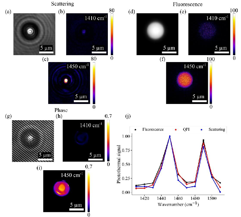
In an innovative breakthrough, researchers have developed a new type of microscope capable of using multiple imaging techniques simultaneously, a game-changer for both material science and biological research. This revolutionary device employs mid-infrared photothermal (MIP) imaging, allowing scientists to choose the best contrast mechanism for their specific samples, whether they are studying cells or polymers.
Introducing the Multi-Contrast Wide-Field MIP Microscope
The research team behind this innovative technology—led by Anooj Thayyil Raveendran and including experts from the Leibniz Institute of Photonic Technology—has designed a multi-contrast imaging platform that utilizes three distinct mechanisms: scattering-based detection, fluorescence modulation, and quantitative phase imaging (QPI). Up until now, various MIP systems have been restricted to a single imaging modality, necessitating the use of different instruments for each. This new design allows researchers to switch between methods depending on their imaging needs, vastly improving flexibility and efficiency.
How Does It Work?
The mid-infrared (MIR) absorption provides high molecular specificity while achieving exceptional spatial resolution. This combination is particularly crucial in identifying various cellular components and materials without labeling them, making it a label-free imaging method. Each method comes with its own benefits: scattering is advantageous for its straightforward setup, fluorescence provides high detail for specific structures, and QPI captures comprehensive thickness information of the sample.
Exploring the Results
The research detailed the system's effectiveness through an array of tests using various samples, including human lung adenocarcinoma cells, mouse lung tissue, and even bacteria. The microscope showed an impressive ability to resolve features down to 376 nm in spatial resolution for scattering, 429 nm for fluorescence, and 904 nm for QPI. Notably, QPI displayed a signal-to-noise ratio (SNR) 26 times higher than that of scattering methods, indicating its superior sensitivity.
The Impact on Biological and Material Research
This multi-contrast microscope could revolutionize how scientists study and diagnose diseases and analyze materials. By facilitating simultaneous imaging of different properties and materials, researchers can gather richer data in less time, potentially leading to quicker diagnoses in medical settings and better understanding of material properties for engineering applications.
A Step Forward in Clinical Applications
Looking forward, the researchers aim to enhance the technological readiness of this wide-field MIP microscope. The ultimate objective is to make it more applicable in clinical settings for rapid screening of bacteria, cells, and even tissue sections—paving the way for a promising future in both medical diagnostics and materials research.
With this advancement, the scientific community is poised to unlock new insights in both biology and material science, exemplifying the dynamic nature of scientific innovation in addressing complex challenges.
For more insights into the intricate workings and potential of this cutting-edge technology, the full study can be referenced from the Leibniz Institute of Photonic Technology.